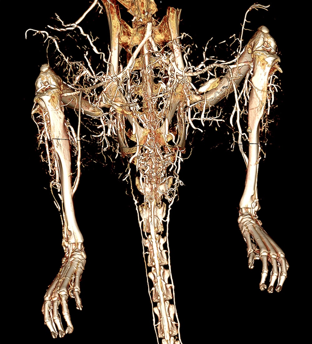
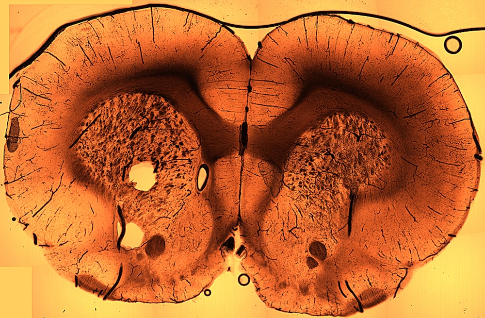
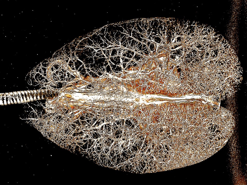
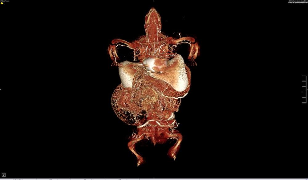
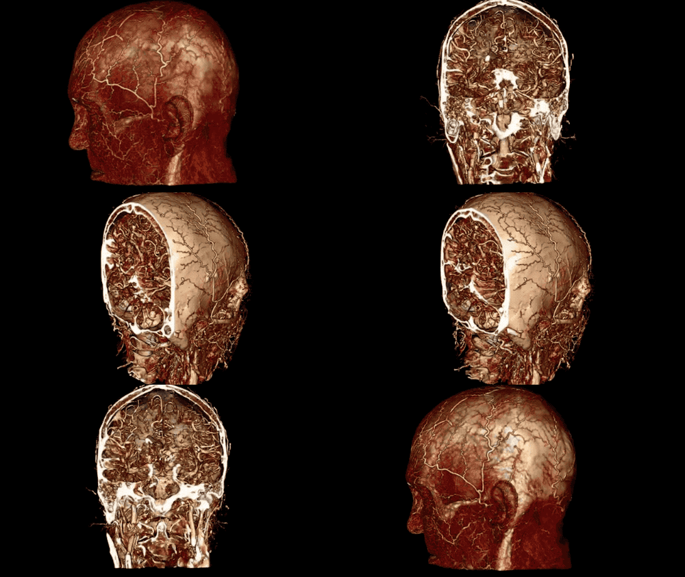
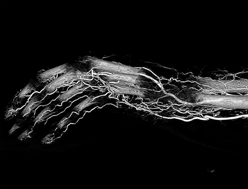
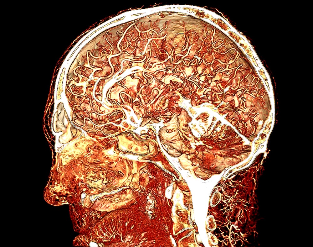
Some say that beauty’s only skin-deep. But one veterinary surgeon and his team look beneath the surface…literally. Scott Echols says that we don’t actually know very much about the anatomy of animals because we haven’t had a way to properly visualize it. But now, with the help of a new imaging technology called BriteVu, researchers have access to a trove of data on animal anatomy.
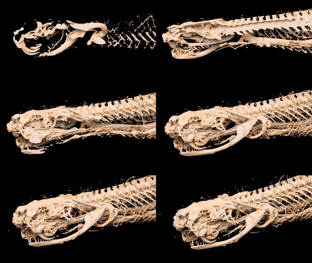
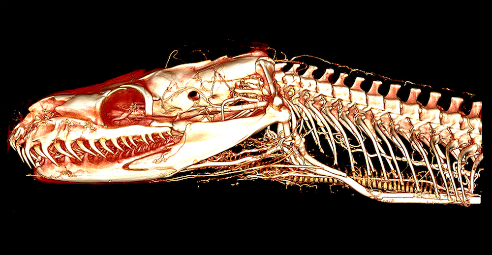
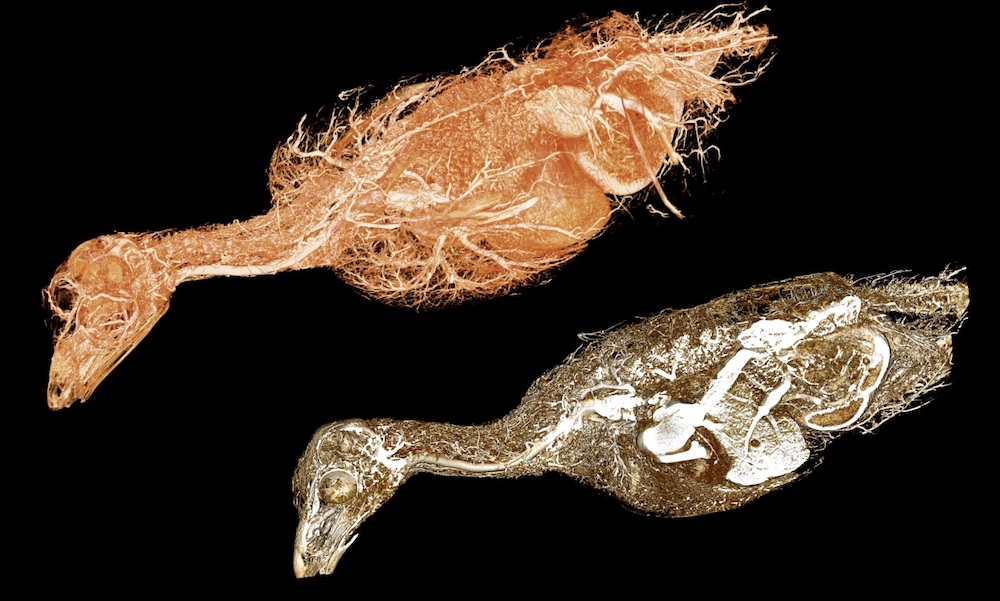
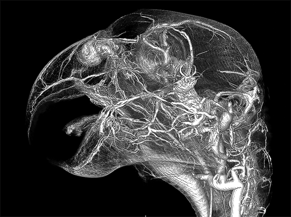
After an animal has been euthanized, researchers inject a compound into its blood vessels that allows them to take a CT scan and create multiple 3D images, which allow researchers to analyze the intricate anatomy that lies below the skin. They can see every blood vessel in the body, from capillaries to the arteries, and everything in between.
“We find things that we never knew existed that were right in front of us all this time,” says Echols. “We can take those animals and we can bring them back to life. They still have a lot more to tell us. Let’s make the most of that information.”
Take a peek at the hidden beauty of animal anatomy:
Credits
Produced by Luke Groskin
Filmed by Dusty Hulet
Music by Audio Network.com
CT Scans and Video provided by Scarlet Imaging, Anatomage, University of Utah,
and the staff & owners of Parrish Creek Veterinary Clinic, Centerville, Utah
Sketches by Ludwig Bojanus, Johannes Sobotta, Herbert Spencer Jennings,
V. Ghetie, Frank E Beddard
Special Thanks to Shane Richins and Robert Groskin.
Meet the Producer
About Luke Groskin
@lgroskinLuke Groskin is Science Friday’s video producer. He’s on a mission to make you love spiders and other odd creatures.