Here’s What A Slice Of Mouse Eye Looks Like
A special imaging technology peers inside a mouse eye, revealing the distinct roles that cells play in maintaining retinal health.

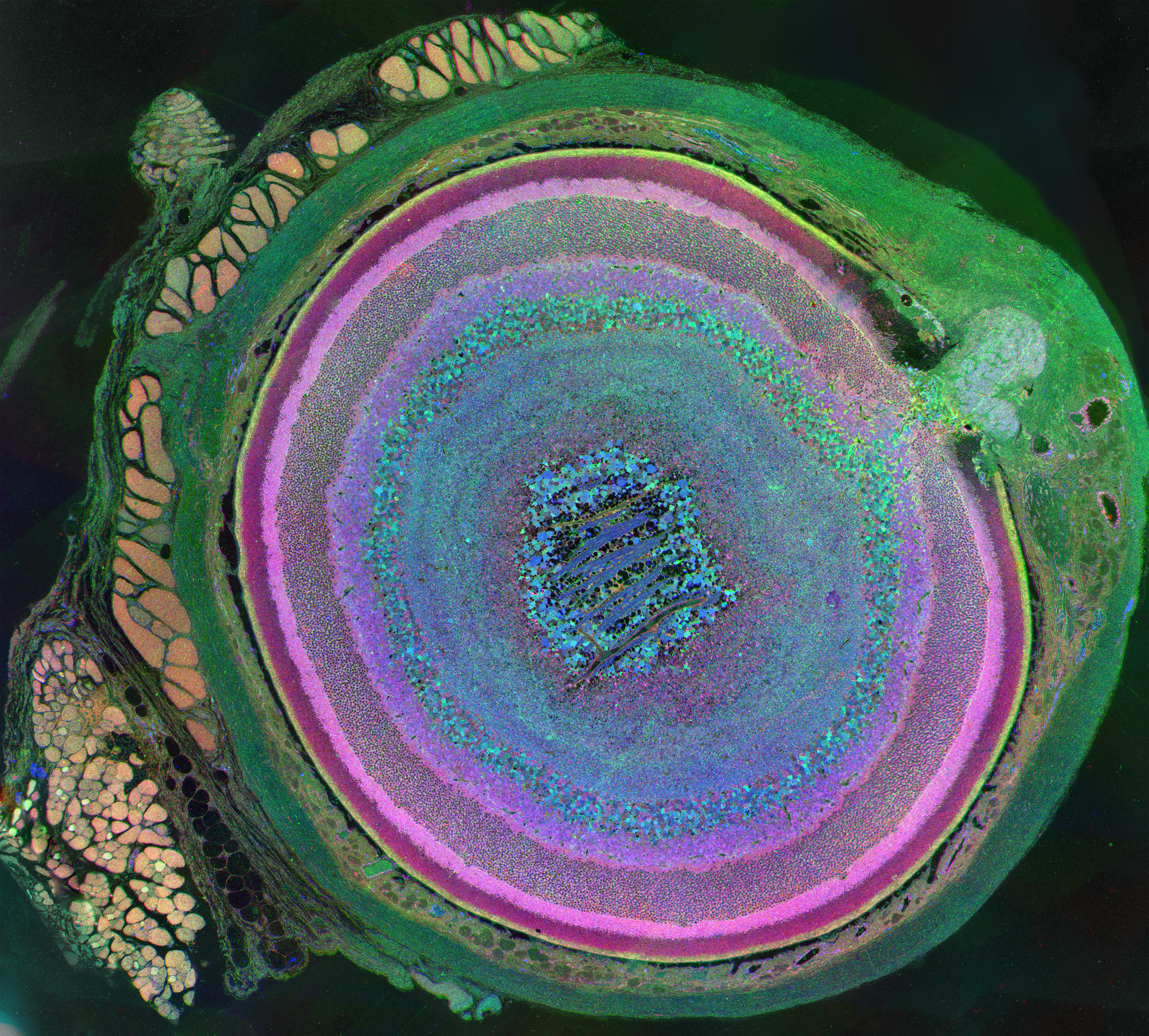
This image of a mouse eye is part of an exhibit of scientific images called "Life, Magnified," currently on display at Washington Dulles International Airport's Gateway Gallery through November. Image by Bryan William Jones and Robert E. Marc, University of Utah
What looks like an expansive crater on some exotic planet is in fact a view into a common mouse’s eye. Each of the vibrant colors reflects the identity and metabolic function of one of the 70-80 types of cells in the mammalian retina.
To create the image, Bryan Jones, a retinal neuroscientist at Utah’s Moran Eye Center, turned to an imaging technique called Computational Molecular Phenotyping (CMP). He first shaved off parts of a mouse eye, revealing layers bit-by-bit—“like licking a gobstopper,” he says. When he reached the halfway mark, he sliced off several microscopically thin layers and applied antibodies that were engineered to attach to particular molecules involved in metabolism.
From those slices, he chose three in which the antibodies had tagged different molecules—taurine (which, among other roles, controls water concentration in cells), glutamine (which provides energy), and glutamate (which helps neurons transmit information)—and used a computer program to scan the slices.
Jones then assigned each tagged molecule a color—red, green, and blue—and sandwiched the resulting scans together. “The colors that emerge are combinations of red, green, and blue signals,” Jones explains, and they reveal information about each cell’s role in metabolism.
For instance, in the composite image, cells carrying more taurine and glutamate are visible in the vivid magenta and pink rings, which represent the inner and outer photoreceptor layers in the retina, where lots of signaling and water regulation take place. The bluish, opaline ganglion cells at the center suggest that glutamate is prominent and that signaling is also occurring.
What the researchers found striking was how the mouse’s retinal cells appeared as distinct, uniformly colored bands, which indicated that each cell had a clearly defined metabolic role. In contrast, when CMP is applied to an aging or diseased human retina, a given retinal layer might appear more mottled. For instance, what appears as a thin yellow layer in the healthy mouse eye “often looks like a brick wall made of different colors” in an aging human eye, says Robert Marc, director of research at the University of Utah’s Moran Eye Center, who developed the CMP technique. That variegation might signal a breakdown in communication between cells, possibly indicating the onset of disease or degeneration in the retina.
Marc and Jones are now using the same imaging technology to track changes in communication between cells in human eyes and to illuminate the differences between healthy and unhealthy retinas. “If we want to be able to effectively intervene with therapies in blinding diseases, then we need to understand what happens to the circuitry in disease,” says Jones.
While the mouse retina gives Marc and Jones a model image of a healthy eye, its complexity suggests there’s still a lot to learn. “When you first make the image, you don’t realize what you’re looking at,” says Jones. “And it slowly dawns on you just how much information is in there.”
Emma Bryce is a freelance science writer whose articles have appeared in publications such as The Guardian and Audubon.