Grade Level
6 - 8
minutes
1- 2 hrs
subject
Life Science
Activity Type:
microorganisms, microscope investigation, taxonomy and classification
There are more than 50,000 different types of microorganisms known to scientists -– and they discover more every year. In a basement laboratory at the University of Pennsylvania, two robotocists have harnessed the sensing, swimming, and swarming abilities of microorganisms to power microscopic robots. Even though their work sounds like the prologue to a dark science fiction film, Ph.D. students Elizabeth Beattie and Denise Wong hope these initial experiments with nano bio-robots will provide a platform for future medical and micro-engineering endeavors.
In this activity, students will learn how to prepare deep well slides for observing two types of microorganisms called Paramecium (a group of protozoa, or single-celled organisms, which move with cilia, so they are called “ciliates”) and Euglena (microorganisms which move with flagella, so they are known as “flagellates”). Students will observe these microorganisms through a microscope, and compare and contrast the physical characteristics of each type of microorganism. Based on their observations and their understanding of flagella and cilia, students will be able to identify which microorganism is the flagellate and which is the ciliate.
Grade Level: 6th – 8th grade
Subject Matter: Life Science, Nanotechnology
National Standards
Cilia — the little hairs that propel a paramecium — flap spontaneously, and will synchronize their movements with neighboring cilia. But scientists have had trouble pinpointing exactly why cilia “do the wave” like this, because cilia are such complicated structures.
Activity Materials
- The following materials can be purchased at any science supply store or online at Carolina Biological Supply Co., http://www.carolina.com:
- At least one microscope
- Medicine droppers or plastic disposable pipettes – one for each student
- Euglena culture (small jar that contains enough for 30 students)
- Paramecium culture (small jar that contains enough for 30 students)
- Deep well slides – one for each student
- Protoslo quieting solution – one small bottle. This solution slows the rapid movement of microorganisms without interfering with them, to help keep them in focus under a microscope.
Other general supplies:
- Toothpicks – one for each student
- Roll of paper towels
- Permanent markers – one for each pair of students
- Paper – enough sheets for all students
- Pencils- one for each student
Vocabulary
Cilia: a group of hair-like structures that assist organisms with locomotion.
Ciliate: an organism that uses cilia for locomotion.
Flagellum: a single hair-like structure that assists an organism with locomotion.
Flagellate: an organism that uses a flagellum for locomotion.
Microorganism: a tiny organism, often made of a single cell, that can be seen only under a microscope.
Paramecium: a group of protozoa, or single-celled organisms. Paramecium move with cilia, so they are called ciliates.
Euglena: a genus of diverse unicellular organisms, some of which have both animal and plant characteristics. (They eat food the way animals do, and can photosynthesize, like plants.) Euglena move with a single flagellum, so they are called flagellates.
What To Do
Note: While all the microorganisms used in this activity are safe for classroom use, be sure to read all safety and care information that comes with your cultures. If students are unfamiliar with using a microscope, plan to introduce basic microscopy and laboratory protocol before teaching this lesson.
- Begin the lesson by having the students watch the SciFri Video, “Dawn of the Cyborg Bacteria” What type of microorganisms were the researchers studying? What type of science tools did they use to observe microorganisms?
- Tell students that they are going to observe the same types of microorganisms (ciliates and flagellates) discussed in the video, and that they will compare the differences and similarities between each microorganism.
- Inform students that it is important to follow proper procedure for preparing a slide. Demonstrate proper slide preparation procedure for them, as outlined below:
a. Hold up the deep well slide and take the cover slip off.
b. Use the dropper or pipette to place five drops of culture in the well of the slide. If there is still space, continue to add culture until the well is almost full.
c. Inform students that the next step is to add two drops of Protoslo to the culture. Ask students to guess what they think Protoslo will do to the microorganisms. What does the name indicate? (Protoslo is a harmless chemical that will slow down microbes without interfering with their characteristic movements, so that the students will be able to observe them through the microscope.)
d. Use a toothpick to gently mix the culture and Protoslo together. (When students begin to look through the microscope, they may find that microorganisms are still moving too quickly to make observations. If so, they should add one or two more drops of Protoslo.)
e. Carefully, replace the cover slip so that there is almost no air trapped in the slide. One technique is to place the cover slip on the edge of the well, and gently slide it completely into place over the well. This might take a few attempts. - Once you have reviewed and demonstrated proper procedure for slide preparation, hand out to students the droppers/pipettes, deep well slides, toothpicks and Protoslo. Depending on class size, you may pass around the jars of cultures, or you may set up them up on a table in a separate area. Have paper towels on hand and keep some culture in reserve in case of spills.
- Divide students into pairs, with one student preparing the Euglena slide and the other preparing the Paramecium slide. Students should use a permanent marker to label the Euglena slide with a small “E” on the edge of the slide, and the Paramecium slide with a small “P.” Inform students that these are the names of two different types of microorganisms.
- Once the slides are prepared, have students use paper and pencils to create a chart with two columns. They should label the first column “Euglena,” and the second column “Paramecium.”
- Have students place the Euglena slide on the microscope stage. Center the slide so that the culture is under the light path.
- Using the low power objective (4x), have students take turns looking through the eyepiece. Students may need assistance in focusing the image.
- Ask the students to record their observations (color, shape, method of locomotion, etc.) under the Euglena column, and to include a sketch of the microorganism.
- To observe the microbes in more detail, have students switch to a higher power objective (10x) and adjust the sharpness of the image with the fine adjustment knob. Have students record any additional observations that they were not able to see at the lower magnification. Warning: If you are using a standard microscope, do not use the 40x objective with the deep well slide. At this point, the objective might be too close to the slide.
- Repeat steps 7-10 for the Paramecium slide.
- Have students compare and contrast their observations. Review the definitions for a flagellate and a ciliate. Can they identify which microorganism is the flagellate and which microorganism is the ciliate? What characteristics do both microorganisms share? What characteristics are different in each? What are some of the advantages to each type of locomotion?
What’s Happening?
Ciliates are microorganisms with small, hair-like projections on their surface called cilia. The cilia can be very numerous, covering the entire surface of many microbes, or several can be fused together to form a bundle. Cilia beat in a coordinated fashion to propel the organism through the water. Flagellates move by beating or twirl single whip-like flagella (longer hair-like appendages, compared to cilia) that extend from their bodies.
Paramecium is a group of slipper-shaped ciliate covered with cilia that live mainly in freshwater environments and feed on yeast and bacteria. (Recently, some new species have been discovered in the oceans.) Paramecia move swiftly and gracefully through the water by the coordinated beating of their cilia. As they swim, they also rotate on their longitudinal axis, rolling like acrobatic airplanes. Each paramecium has a depression called an oral groove along one of its sides. The water flow created by the coordinated beating of the cilia forces food into this groove. Therefore, the cilia serve not only as a means of locomotion, but also as a feeding mechanism.
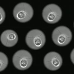
Euglena is a very common green flagellate that can be observed by the unaided eye when millions of them gather to form a green film on the surface of a pond. Euglena has a single whip-like structure located at one end of its body that pulls it through the water. Euglena also has a flexible cell wall that allows it to twist and turn in a characteristic maneuver known as euglenoid movement. Besides exhibiting the animal characteristic of locomotion, some Euglena exhibit plant characteristics as well. The green pigment observed on euglena is chlorophyll, which it uses to convert light into food by photosynthesis. It lives in fresh and salt water.
Topics for Science Class Discussion
- What do Euglena and/or Paramecium need in order to survive? How would you maintain the lives of these microorganisms as classroom pets?
- Why did we use a deep well slide for this investigation and not a flat microscope slide and slip cover?
- Compare the movements of ciliates and flagellates to the movement of humans in water. How are they different or similar? Which form of locomotion provides better maneuverability and speed?
Extended Activities and Links
Extend the activity by collecting samples from a local pond or lake. Make comparisons between the microorganism from a laboratory and those collected in the field.
Have students observe microorganisms that do not use flagella or cilia for locomotion by researching them online. Students can create a poster board presentation outlining the advantages or disadvantages between each method of locomotion.
Working individually or in groups, have the students use what they have learned about microorganisms and locomotion to design their own microorganism. Let students use a variety of media to portray their completed organisms; e.g., drawing pencils, pens, charcoal, paint, clay for models.
Learn more about microorganisms through this interactive website:
http://www.childrensuniversity.manchester.ac.uk/interactives/science/microorganisms/whatandwhere.asp
Learn more about the group of microorganisms called bacteria by visiting:
http://ilovebacteria.com/microbes.htm.
See more amazing microscopic images and learn about the art of “cinemicroscopy” by watching the SciFri Video “Microscopic Movie Stars”:
This lesson plan was created by the New York Hall of Science in collaboration with Science Friday.

The New York Hall of Science is a science museum located in the New York City borough of Queens. NYSCI is New York City’s only hands-on science and technology center, with more than 400 hands-on exhibits explore biology, chemistry, and physics.